Only at RIT: Looking at Food in a New Way
PHPS 321 – Photography Using a Light Microscope
by Professor Michael Peres
The High Magnification Photography discussed what the final project of the High Magnification Photography class should be in week seven, and then in week eight, and then in week nine and then …..
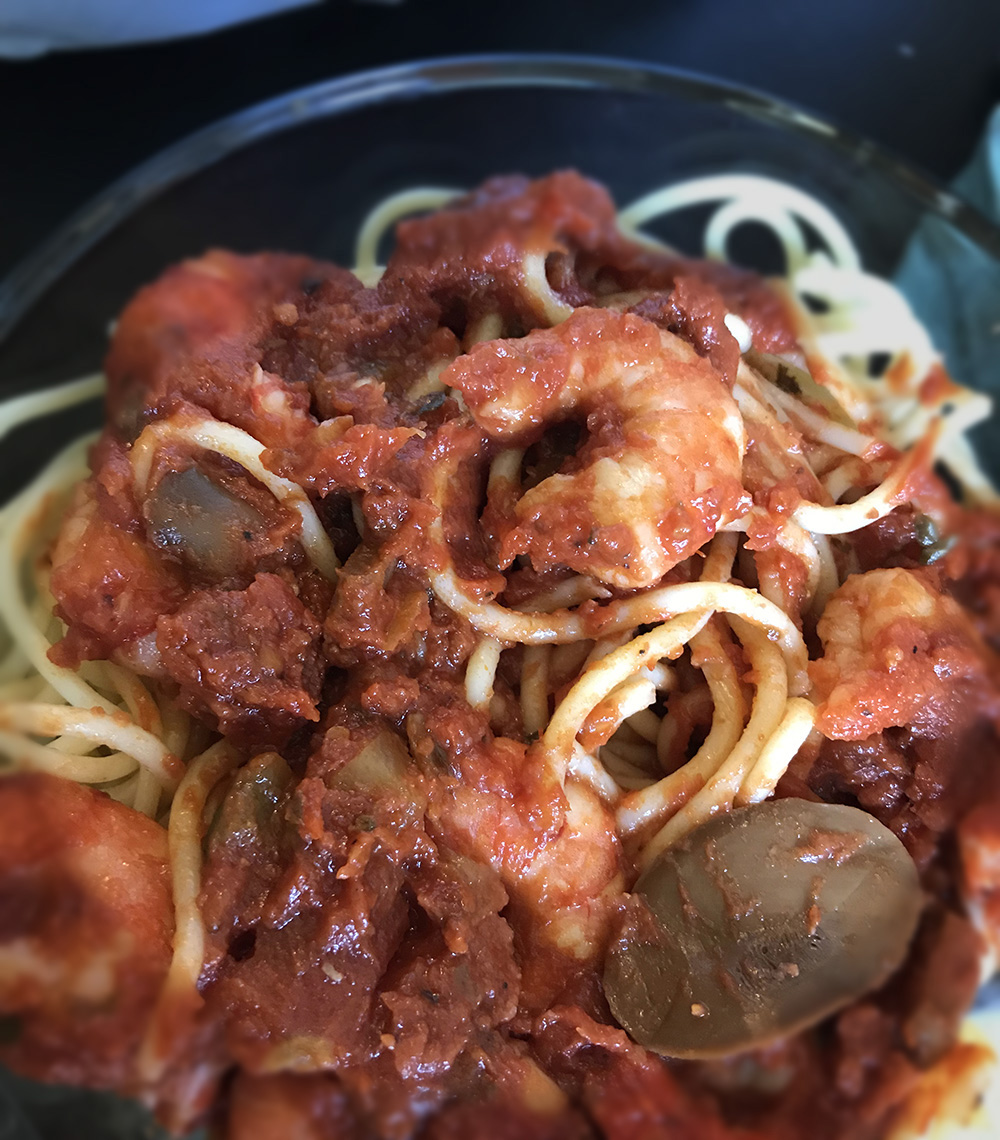
Really, it was not that complicated but it took some time for the students to agree on a theme. In the end, they decided to look at ingredients of food. Each student was required to produce 10 photomicrographs exploring the ingredients of some food they were curious about. One student wanted to photograph the ingredients from the recipe of her grandfather’s favorite pound cake for example and another loved chicken fajitas.
Photographing large objects using a light microscope is a challenging process. Separate from the influences created by the sample, images produced from a microscope have virtually no depth of field. Samples have topography and surface characteristics that need to be made visible using various techniques and illuminations. There are a lot of issues. Shared below are four examples from each student. We hope will enjoy the visual solutions to a number of challenges each of the students resolved.
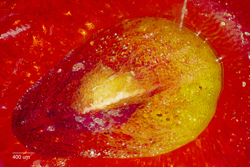
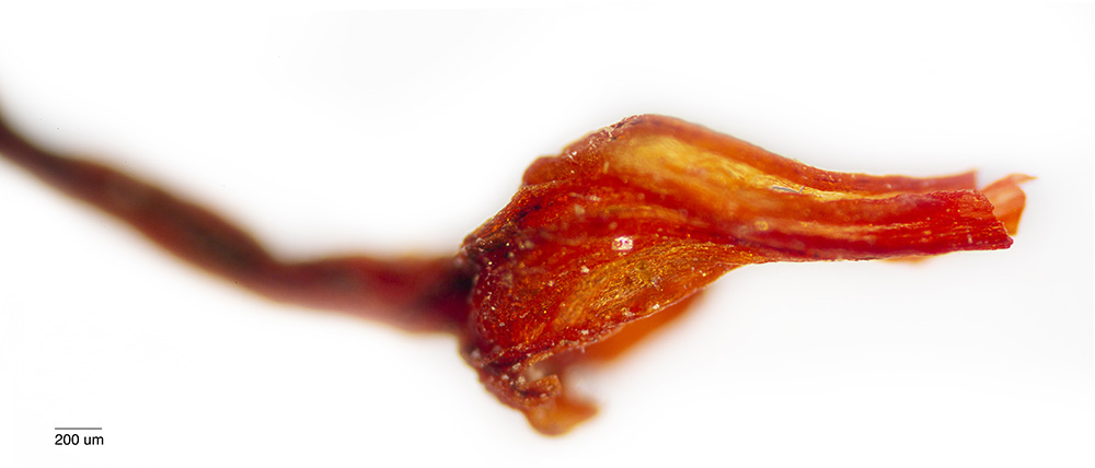
Ethan Whitecotton photographed tea leaves and fruit

A blueberry seed
A strawberry seed
A tea leaf

The stem of a blueberry
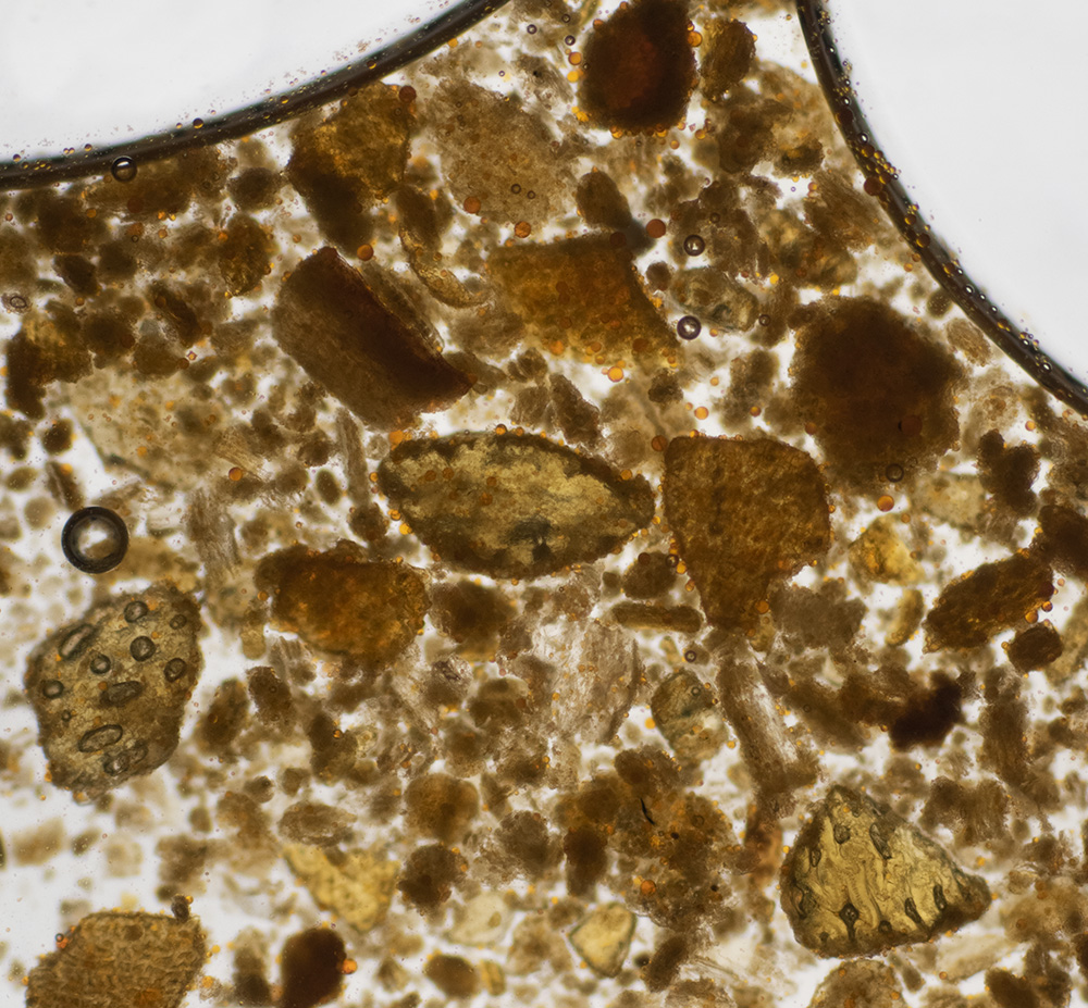
Lydia Dye photographed spices.
Cayanne Pepper

Coconut Oil

Honey
Oregano
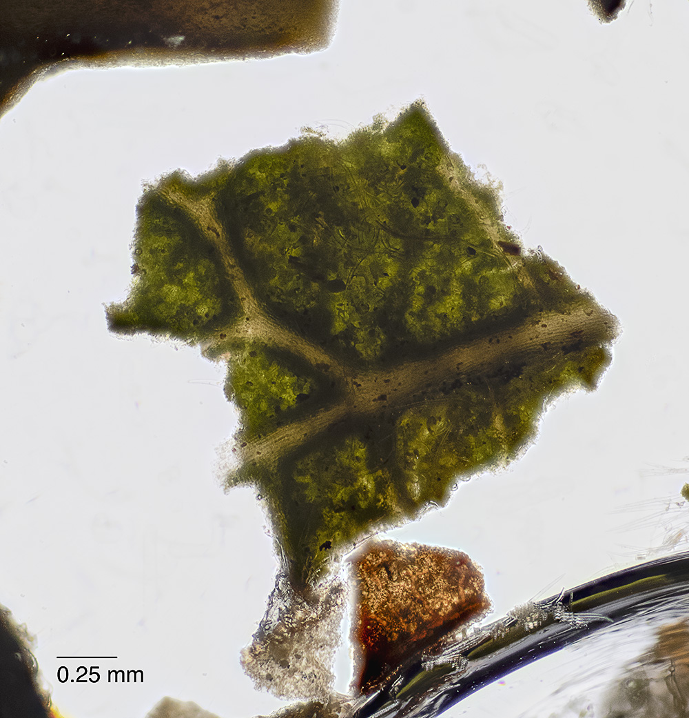
Kristen Hanley photographed the ingredients from chicken fajitas
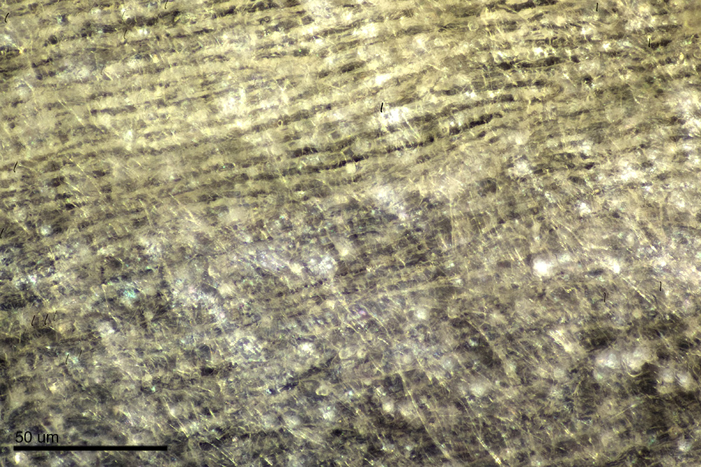
Garlic Skin

Pepper Seed

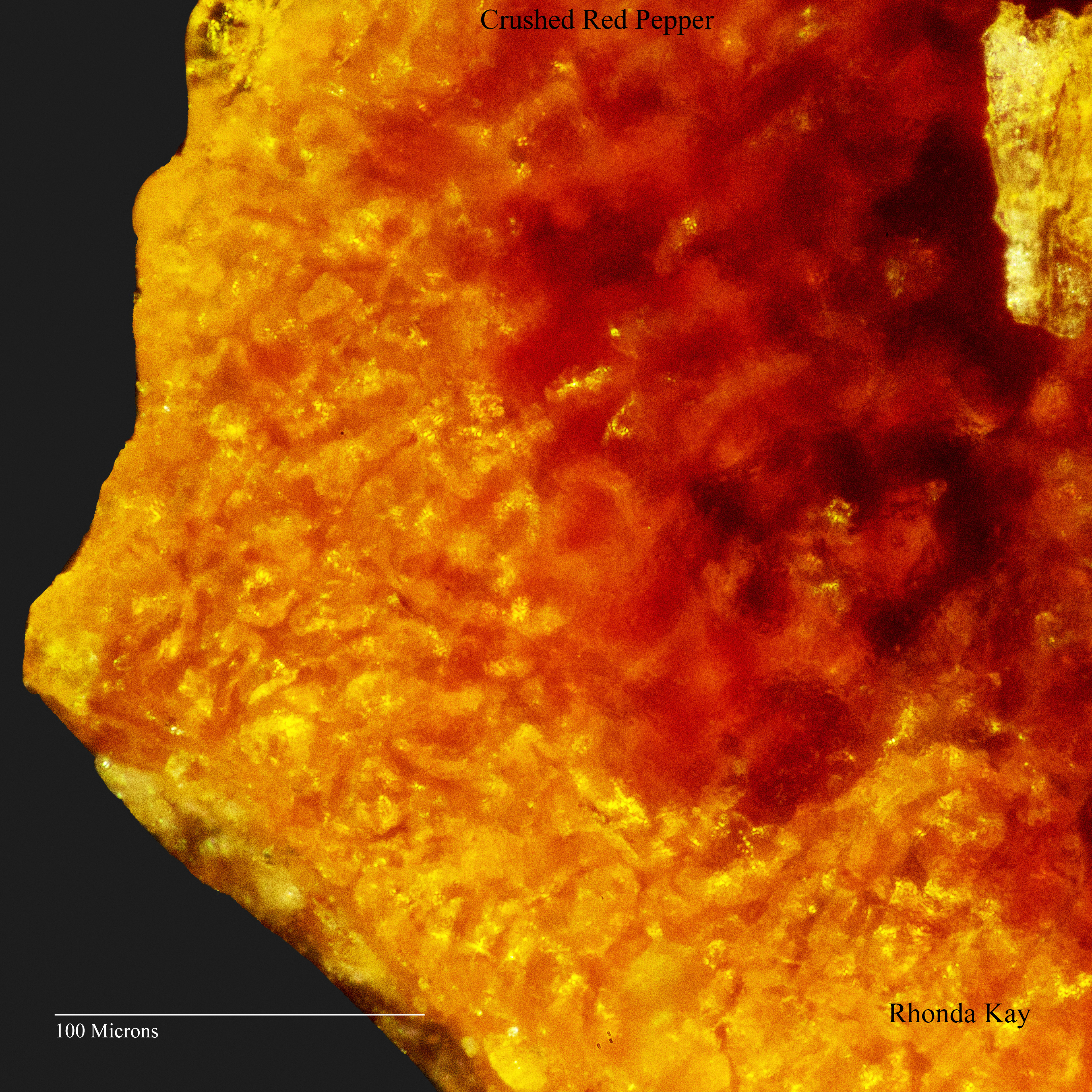
Red Pepper Flake
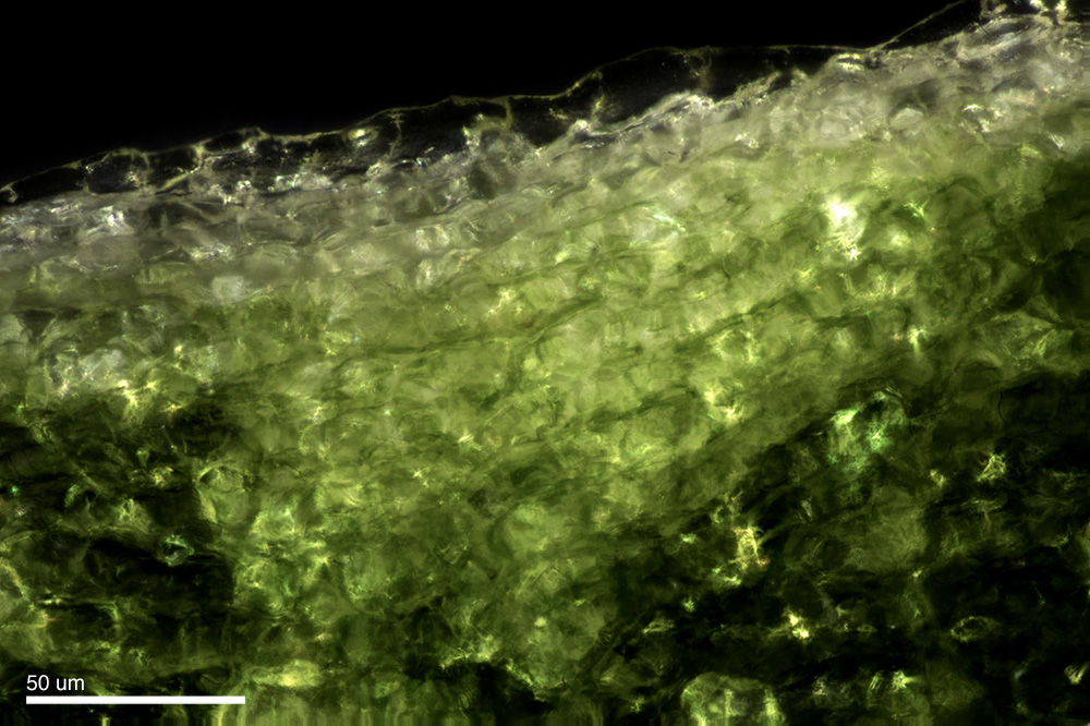
Green Pepper
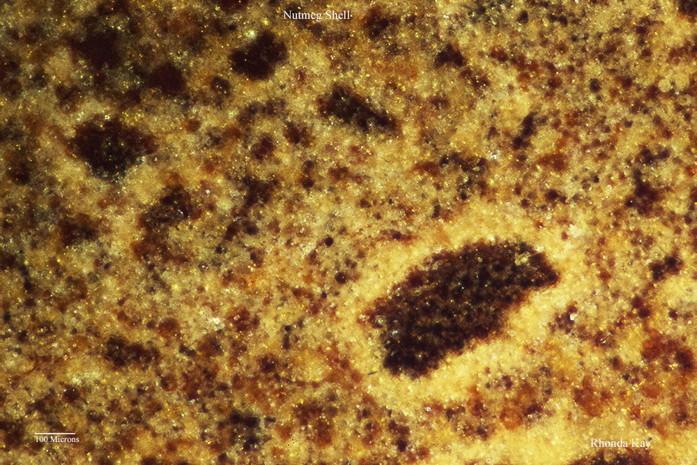
Rhonda Kay ultimately ended up photographing spices. The ingredients used in pound cake such as oils, produced really nothing that was visually strong enough to pursue.


About Michael Peres
Michael Peres is an award winning photo-educator, author, and science photographer. Peres is a professor of biomedical photographic communications and teaches photomicrography, biomedical photography, and other related applications of photography in science at Rochester Institute of Technology.
Peres has enjoyed a varied photographic career which, began in 1973. He has been actively publishing most of his career and recently wrote Laboratory Imaging and Photography published by Focal Press in 2016 as well the Focal Encyclopedia of Photography – 4th Edition. He is also a co-author of Michael Photographs a Snowflake, children’s book published by Fossil Press also in 2016.
Peres was a 2003 RIT outstanding faculty award winner and he has been twice awarded the Gitner prize presented by the RIT College of Imaging Arts & Sciences for outstanding achievement in the graphic arts. In 2007, Peres was awarded the Schmidt medal by the BioCommunications Association for lifetime achievements in the field of bio-communications. Michael holds a master’s degree in instructional technology and bachelor’s degrees in biology and biomedical photographic communications. He is also a registered biological photographer.