From the Classroom: the Photographic Sciences Capstone Experience
Photo Sciences Capstone experiences are Application Oriented by Christye Sisson
Greetings from the depths of the Fall semester at RIT! I’m Christye Sisson, program director of the Photographic Sciences at RIT Photo. We are all looking forward to Thanksgiving Break to take a little breather and spend time with friends, family and food. Our seniors have been working hard this semester in the Photo Sciences Capstone proposals prior to starting production in the spring semesters’ project phase in Capstone II.
The capstone class started 6 years ago in 2013, when RIT converted to semesters from its traditional quarter curriculum. As part of the conversion, the University wanted a way for undergraduate students to have a culminating experience for each degree program. The capstone class is a way for students to spend one or two semesters on an original self directed project specific to their degree that combines elements of their educational experiences in their programs. Our Photo Sciences Capstone experience is very application oriented and students look to solve practical problems while also applying what they’ve learned in the classroom and on coop.
The capstone experience can be self-originated, in coordination with a faculty member engaged in applied research, or as a partnership with industry looking to solve specific research problems. By approaching the experience in a variety of ways, we give the opportunity to the student to come up with their own project, while also supporting industry or research-led initiatives. In all cases, the students work directly with a faculty member, usually in the Photo Sciences but often in conjunction with professors from other disciplines, to advise them through the process. The students must learn proposal writing, project management, asset management, resource allocation, budgeting, and problem solving as they navigate their way through their capstone.
Past projects have included students who created educational 3D models and animations of a camera system including optics, and with working animated moving parts to control virtual optical and image outcomes. In another project, a student worked to create a multi-spectral test target for use in calibration of IR/UV camera applications. Another student rekindled the Victorian practice of diatom arrangement to create stunning kaleidoscopes that would fit inside a period printed on a piece of paper. Still another built a sensory substitution device that a visually impaired person could use to navigate their environment, translating objects into sound in a three-dimensional space.
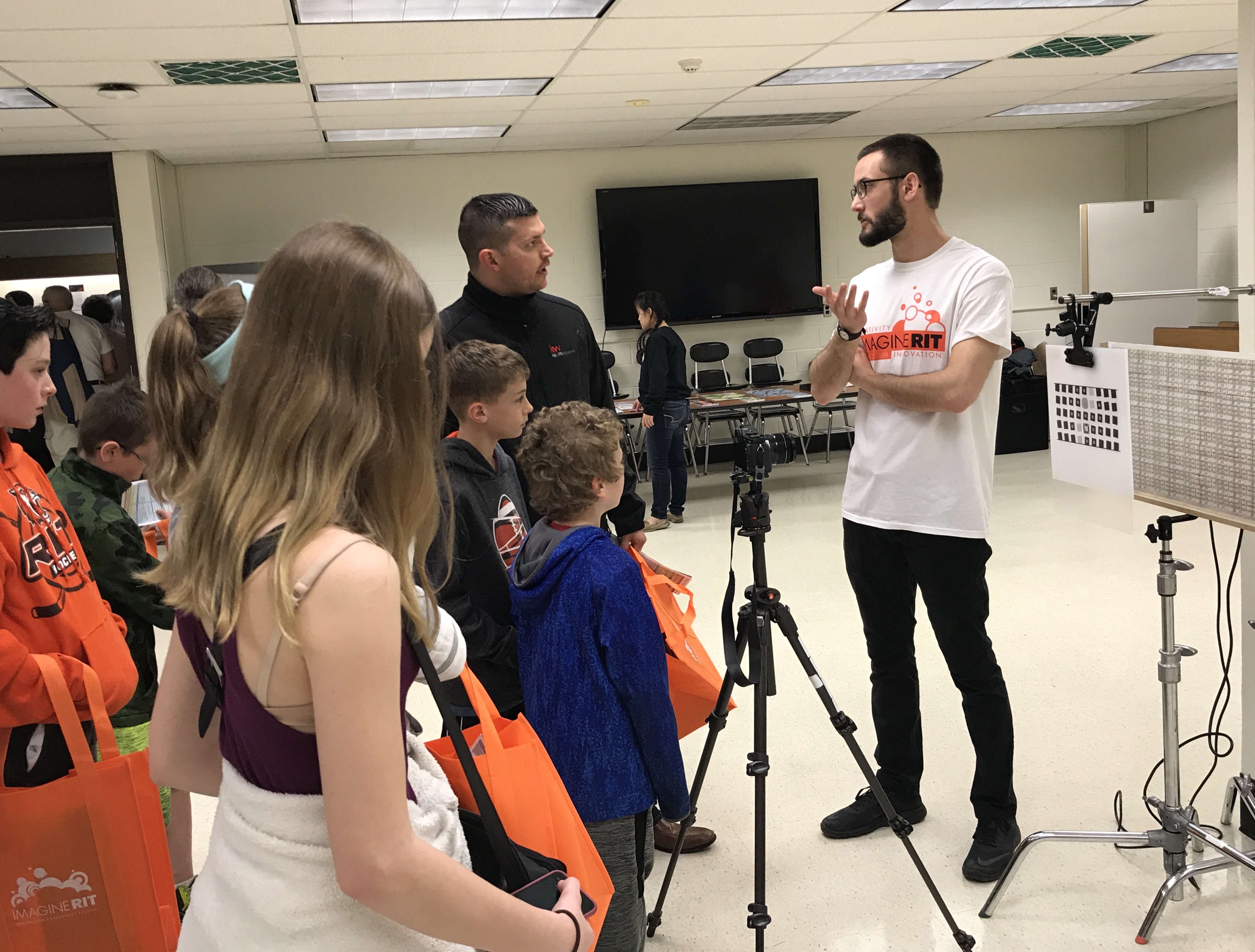
Caption: Photo Sciences student Ian Longton explaining his targets for Near-Infrared Image Quality Testing at ImagineRIT.
We’ve had many students generate educational materials as part of their projects, using stunning photography to communicate scientific concepts to a wide variety of audiences. Sometimes these are in the form of traditional educational materials like web sites, posters and videos, and other times they can be in the form of a children’s book on the hidden world of a home aquarium.
Industry partnerships make up a growing portion of our capstone projects, with one of the most prominent being the Facebook 360˚ project. Facebook generously gifted us with all of the parts and resources to build their open source, 360 degree still and video camera, while helping them to solve image quality and color management issues along the way. The National Science Foundation’s sponsorship has fostered a collaboration between RIT’s College of Engineering’s Multidisciplinary Senior Design Project and our students to work on building a low-cost retinal camera for the last three years. And the Food and Drug Administration, in conjunction with the International Color Consortium, has partnered with our faculty and students to devise a color management protocol for retinal cameras, devices that capture images of the retina.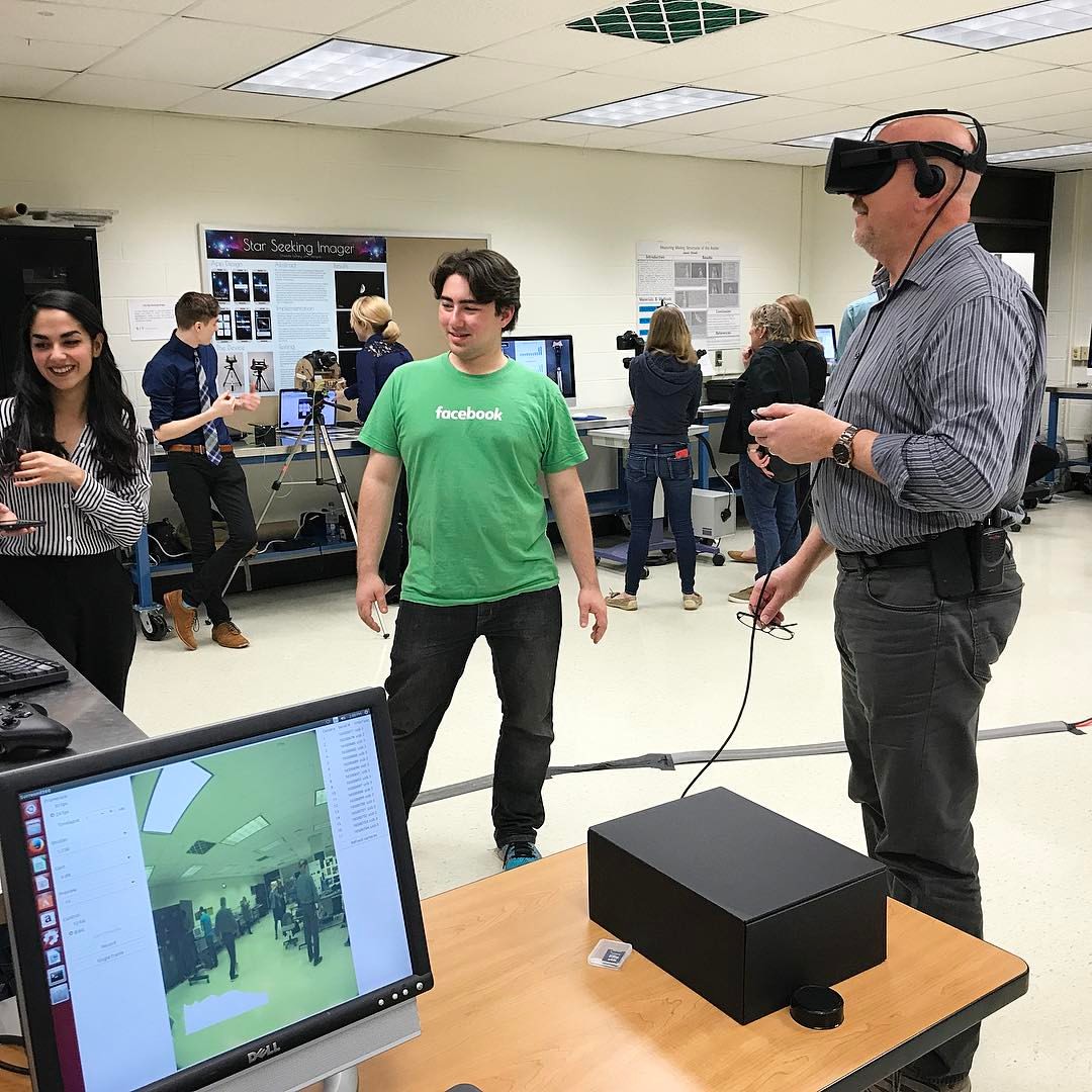
Caption: RIT Photographic Facilities Coordinator David Walter tests out the Facebook 360˚ environment with Nick Franco and Madison Zic.
We look forward to this years’ projects as student’s finalize their plans and tease out the difference between what is possible and what is practical. Inevitably, even though there are (always) issues that come up along the way, our students produce a remarkable body of work that they, and we, are proud of. Just last year, our students presented their capstone work at three separate professional conferences, made possible through the generosity of alumni support.
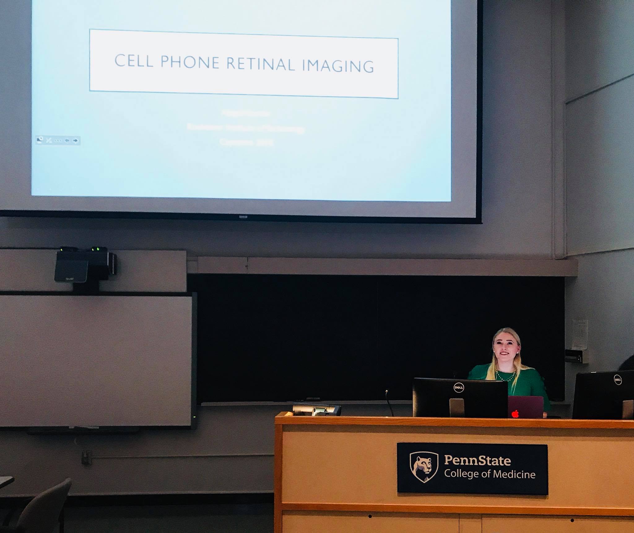
Caption: Abby Semler presented her project on Cell Phone Retinal Imaging Quality Assessment to the Imaging Track of the 10th Annual Update for Ophthalmic Medical Personnel in Hershey, PA.
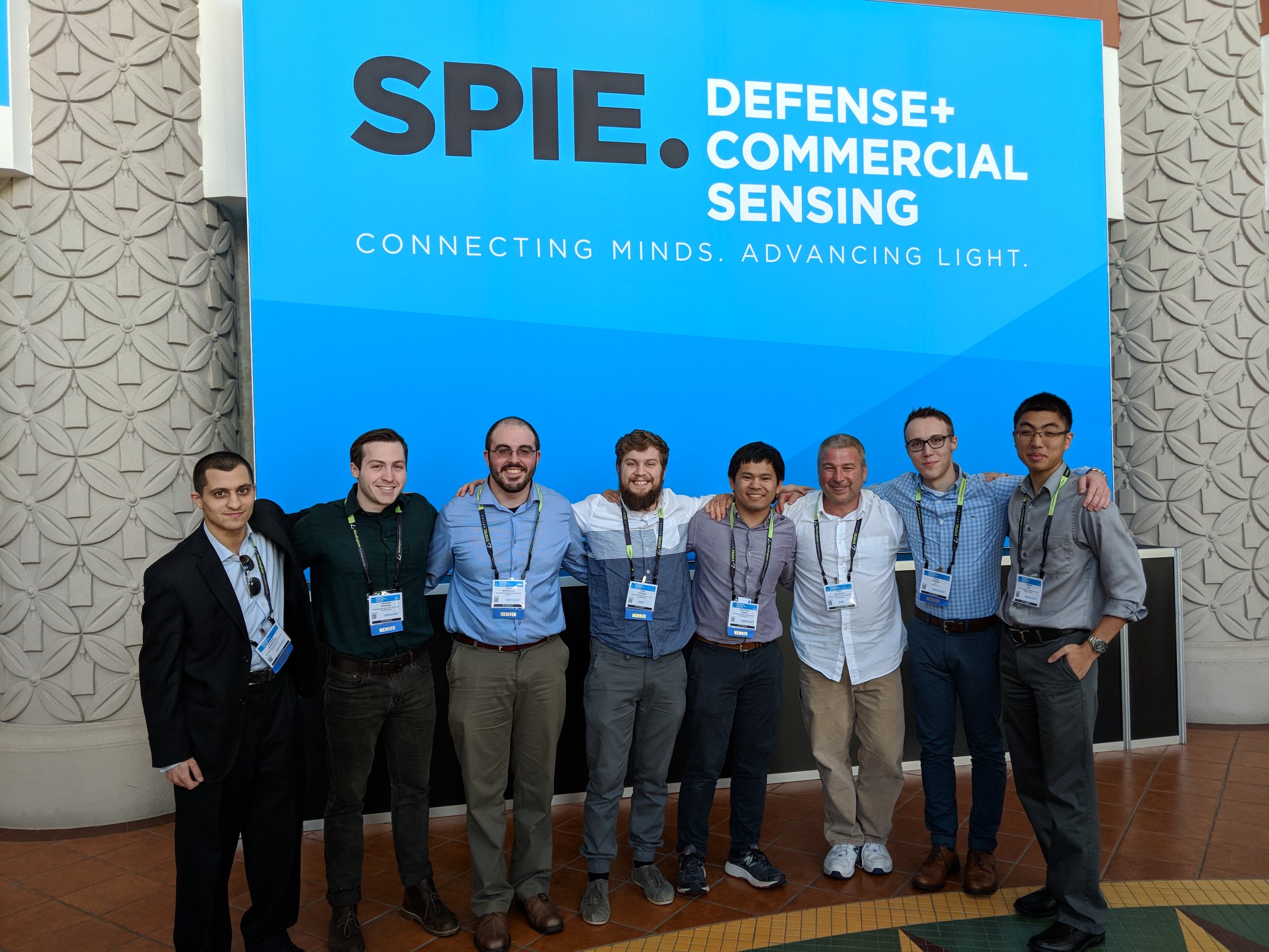
Caption: Geoffrey Sasaki (fourth from right) presented his work, “An exploration of vicarious and in-scene calibration techniques for small unmanned aircraft systems” at SPIE’s Defense and Commercial Sensing Conference in Orlando, Florida. His work was a part of the session on creating reliable image data on UAV’s. Geoffrey also worked with Carl Salvaggio (third from right) from RIT’s Imaging Science department, as his advisor.
Check back in with us in the Spring to see the final result of this year’s student Capstone projects – you can follow our activities on Facebook, RIT Photographic Sciences (https://www.facebook.com/search/top/?q=rit%20-%20photographic%20sciences). And don’t hesitate to contact me at cpspph@rit.edu with any questions you may have. This Thanksgiving, one of the many things I’m grateful to be surrounded by our amazing students, and am so lucky to get to watch over their time with us as they reach and exceed their potential. Happy Thanksgiving!
About the Author:
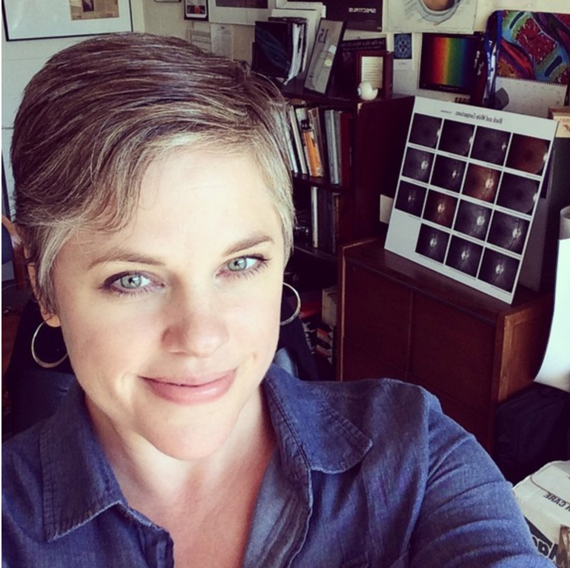
Christye Sisson joined the Biomedical Photographic Communications department at RIT in 1997 after working as an ophthalmic photographer in Boston, Massachusetts and in Rochester, N.Y. Christye holds a Master’s Degree in Information Technology, a Bachelor of Science in Biomedical Photographic Communications, and is a Certified Retinal Angiographer. She was appointed chair of the Photographic Sciences department (formerly Biomedical Photographic Communications and Imaging and Photographic Technology programs) in 2010. Christye is an Associate Professor, and teaches various courses including Ophthalmic Photography I and II, Vision and Perception in Imaging, and Careers and Professional Practices courses for the Photographic Sciences department.
Christye holds a Visiting Faculty appointment at the Flaum Eye Institute, part of the Department of Ophthalmology at the University of Rochester Medical Center. She is involved with a number of image quality and color in fundus imaging projects, including Tele-I-Health, a diabetic retinopathy screening program. Christye is also the project director of the Color Eye Model project, a sub-group of the Medical Imaging Working Group with the International Color Consortium (ICC), co-founded by the FDA and ICC.
Christye is active in the Ophthalmic Photographer’s Society and has hosted the OPS’s CRA (Certified Retinal Angiographer) Exam, acting as both site coordinator and examiner. She is a past member of the OPS Board of Certification, which oversees the certification and administration of the Certified Retinal Angiographer’s exam and activities. Christye oversees all aspects of the ophthalmic activities and equipment that the department maintains, and is also a faculty advisor for the Photographic Sciences Club.